Renal imaging
Biomedical Imaging Laboratory
Chronic Kidney Disease
Chronic Kidney Disease (CKD) is a major social health problem with high prevalence of asymptomatic CKD in apparently healthy subjects in Spain, especially in older, obese and hypertensive patients.
There are some techniques used in the clinical routine to detect the functional changes, however the evaluation of kidney microvasculature remains a challenge.
The main purpose of our research is to provide a non-invasive multiparametric imaging technique to detect functional changes and characterize the renal disease.
We are active members of COST (European Cooperation in Science and Technology) Action PARENCHIMA (CA16103), with the aim of developing and standardizing magnetic resonance imaging biomarkers for chronic kidney disease.

Do you need us to help you?
Contact us and request more information.
Renal magnetic resonance imaging
Renal perfusion
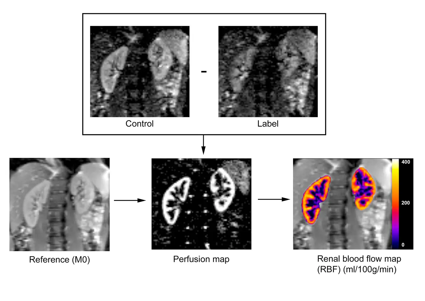
Arterial Spin Labeling (ASL) is a MR imaging technique which employs magnetically labeled arterial blood water as an endogenous tracer to allow quantification of tissue perfusion. Perfusion is calculated as the difference between non-labelled (control) images and labeled images.
Renal blood flow (RBF) can be quantified using a mathematical model which transforms the perfusion signal into physiological units (ml/min/100g).

Figure 1. Arterial Spin Labeling (ASL) overview in which control and label images are acquired. A difference image is obtained for each control-label pair. Multiple ASL pairs are averaged to obtain the mean perfusion map. Renal blood flow (ml/min/100g) is calculated by applying a mathematical model to the mean perfusion image and to the reference image (M0).
Vídeo 1. An example of the ASL technique to measure perfusion in a representative volunteer. In this case, PCASL labeling strategy with an SE-EPI readout were employed.
We have implemented two ASL labeling schemes: Flow-sensitive Alternating Inversion Recovery (FAIR) Pulsed ASL (PASL) and Pseudo continuous ASL (PCASL).
For imaging, we commonly use a balanced steady-state free precession (bSSFP) readout module or a Spin Echo – Echo Planar Imaging (SE-EPI).
Renal Diffusion
Intra-voxel Incoherent Motion (IVIM) technique allows the characterization of microscopic movement of water molecules in the tissue. In diffusion, several images are acquired using different b-values (diffusion gradients).
The diffusion coefficient D, the pseudo-diffusion coefficient D* and the perfusion fraction (PF) are estimated by applying a bi-exponential fitting to the acquired set of images.

Figure 2. a) Diffusion images acquired for the different gradients (b-values). b) An example of bi-exponential fitting to IVIM equation. The signal was measured in the cortex (blue box) for each b-value. On the right, IVIM maps: c) diffusion coefficient D (10-3 mm2/s), d) pseudo-diffusion coefficient D* (10-3 mm2/s) , and e) perfusion fraction (PF) in %.
Clinical applications of multiparametric MRI
These techniques have been employed to measure perfusion in a study with diabetic patients and to measure diffusion and perfusion in transplanted kidneys, showing promising results and motivating to the further development and optimization of the techniques.
Diabetic nephropathy
Diabetic nephropathy (DN) is a microvascular complication of diabetes mellitus (DM) and a leading cause of end-stage renal disease. Diabetic patients are at high risk for developing CKD but clinically evidence of renal lesion is not seen until they have suffered advanced damage.
In this study, we have shown that ASL is able to detect hemodynamic changes in diabetic patients, from early to advanced stages of CKD. RBF maps of type 2 diabetes mellitus patients shown a reduction of 28% in cortical renal blood flow compared with healthy control subjects.
"Arterial spin labeling MRI is able to detect early hemodynamic changes in diabetic nephropathy"
ASL may help to detect patients at high risk of developing diabetic nephropathy.

Figure 3. Renal blood flow maps in a healthy control (A) and in a diabetic patient (B). Color scale represents renal blood flow in ml/min/100g.
Renal allograft
Renal transplantation is the therapy of choice for patients with stage five of CKD. One of the main factors contributing to the good evaluation of renal function is maintaining an adequate and stable renal perfusion. In fact, ischemia related to transplant surgery contributes to initial renal dysfunction in renal allograft and continues to affect renal function.
Renal allograft perfusion and diffusion has been studied in a group of patients with stable renal allograft function (eGFR > 50 ml/min/1.73m2

Figure 4. Diffusion and perfusion maps of a representative transplanted patient (from left to right: D coefficient (10-3 mm2/s), D* coefficient (10-3 mm2/s), perfusion fraction (FP) (%), and renal blood flow map (RBF in mL/min/100g).


