Interventional Radiology Unit
"Interventional Radiology offers effective treatments as an alternative to open surgeries, with less discomfort and faster recovery."
DR. ANTONIO MARTÍNEZ SPECIALIST. INTERVENTIONAL RADIOLOGY UNIT

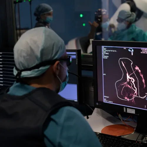
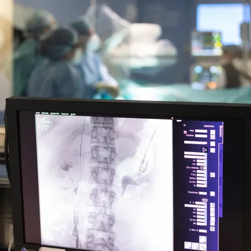
Interventional Radiology is a medical subspecialty that uses advanced imaging techniques —such as fluoroscopy, computed tomography, and ultrasound— to perform diagnostic and therapeutic procedures in a minimally invasive way.
With the help of real-time imaging, specialists insert very fine and precise instruments into the body to treat various diseases without the need for open surgery. Thanks to this precision, procedures such as targeted biopsies, drainages, embolizations, and treatments for vascular diseases or tumors can be performed, among others.
The Interventional Radiology Unit at Clínica Universidad de Navarra is equipped with the most advanced technology and a highly experienced medical team. In addition, it works closely with other departments such as Oncology, Surgery, Urology, Gynecology, Gastroenterology, Pulmonology, Anesthesiology, and Emergency Medicine, ensuring a comprehensive and coordinated approach for each patient, tailored to their specific needs.

A less invasive alternative, more beneficial to the patient

Less
invasiveness
Avoids open surgeries by using small, image-guided punctures

More
security
Reduces risk of complications compared to traditional surgery

Faster
recovery
It reduces pain and allows the patient to return to normal activities within 24-48 hours.

Shorter
hospital stay
In many cases, it allows discharge in less time or even on the same day
Specialized units for a better attention
IN NAVARRA AND MADRID
1. Percutaneous Biopsies
Percutaneous biopsy is a fundamental procedure in the diagnosis of both malignant and benign lesions in various locations. Using advanced image guidance, tissue samples are obtained with minimal morbidity.
Types of biopsies:
- Ultrasound-guided biopsy (breast, thyroid, lymph nodes)
- CT-guided / C-arm–guided biopsy (hepatic, renal, pulmonary, bone lesions)
- MRI-guided biopsy
Indications: Characterization of indeterminate masses, suspicion of malignancy, diagnostic confirmation prior to definitive treatment.
2. Percutaneous Transhepatic Cholangiography (PTC)
Diagnostic procedure that allows visualization of the intrahepatic and extrahepatic bile ducts through percutaneous hepatic puncture guided by ultrasound or CT.
Indications: Obstructive jaundice, evaluation of the biliary tree, palliative biliary drainage in selected cases.
3. Discography and Spine Studies
Diagnostic procedures for detailed evaluation of disc pathology and structures of the spinal canal.
1. Selective Digital Angiography
Digital subtraction angiography is the reference standard for the evaluation of arterial and venous vasculature.
Types:
- Cerebral angiography (arterial and venous)
- Thoracic angiography (pulmonary, aortic, coronary)
- Abdominal angiography (hepatic, renal, splanchnic)
- Peripheral angiography (limbs, abdominal aorta)
Indications: Evaluation of stenosis, occlusions, fibromuscular dysplasia, vascular malformations, suspected aortic lesions.
2. Venography and Lymphangiography
Specialized studies of the venous vasculature and the lymphatic system.
1. Diagnostic and Therapeutic Nerve Blocks
Spinal blocks:
- Translaminar epidural block
- Transforaminal epidural block
- Caudal epidural block
- Medial branch facet block (diagnostic blocks)
- Facet neurolysis by radiofrequency
Visceral blocks:
- Celiac plexus block
- Superior hypogastric plexus block
- Ganglion impar block
Peripheral blocks:
- Intercostal blocks
- Peripheral nerve blocks
- Peripheral nerve neurolysis by cryoablation
- Joint blocks (hip, knee, shoulder, ankle)
2. Joint Injections
Image-guided intra-articular injection procedures for the treatment of degenerative, inflammatory, and posttraumatic joint pain.
Treatable joints:
- Cervical, thoracic, and lumbar facet joints
- Sacroiliac joint
- Shoulder (glenohumeral, subacromial, acromioclavicular)
- Elbow
- Hip
- Knee
- Ankle
3. Joint Embolization
Minimally invasive procedure consisting of selective occlusion of hypervascular synovial arteries using microparticles or embolic spheres, indicated for pain and inflammation control in refractory joint disease.
- Embolization of hypervascular synovium in hemophilia (recurrent hemarthrosis)
- Refractory inflammatory arthritis (rheumatoid arthritis, spondyloarthritis)
- Advanced osteoarthritis with persistent hypervascular synovitis
- Posttraumatic sequelae with pathological vascularization
Indications: Patients not eligible for major surgery, failure of conservative and/or percutaneous treatment, joint preservation in young patients.
4. Trigger Point Injections
Ultrasound-guided injections of local anesthetic with or without corticosteroids for the treatment of chronic myofascial pain.
5. Vertebroplasty and Kyphoplasty
Minimally invasive procedures for the treatment of pain in osteoporotic or pathological vertebral body fractures.
- Percutaneous vertebroplasty
- Balloon kyphoplasty
Indications: Painful osteoporotic vertebral fractures, pathological fractures secondary to metastases, multiple myeloma.
6. Drug Infusion Systems
Placement of neuraxial infusion systems for the treatment of pain refractory to other therapeutic modalities.
1. Thermal Tumor Ablation
Cryoablation:
Controlled freezing of tumors with freeze–thaw cycles that selectively destroy malignant tissue. Particularly useful in renal, pulmonary, and bone tumors.
Radiofrequency Ablation (RFA):
Generation of heat by radiofrequency to induce coagulative necrosis of malignant lesions, particularly indicated in hepatocellular carcinoma, liver metastases, and renal tumors.
Microwave Ablation:
Microwave ablation is a minimally invasive thermoablative technique that destroys tumor tissue through heat generated by a high-frequency electromagnetic field. Particularly indicated in hepatocellular carcinoma, liver metastases, and renal tumors.
Indications: Tumors <4 cm in diameter, patients not eligible for surgery, bridge therapy to transplantation or hepatic resection.
2. Transarterial Chemoembolization (TACE)
TACE is a locoregional procedure that combines selective administration of chemotherapeutic agents with subsequent arterial embolization, maximizing tumor exposure to chemotherapy and inducing ischemic necrosis.
Technical variants:
- Conventional TACE with lipiodol
- Drug-eluting TACE (resin beads or drug-loaded spheres)
- Selective or superselective TACE
Indications: Intermediate to advanced hepatocellular carcinoma without portal vein thrombosis, cholangiocarcinomas, liver metastases.
Advantages: Excellent local control, possibility of multiple treatments, better tolerance in multifocal disease.
3. Radioembolization (TARE-Y90)
Radioembolization, also known as selective internal radiation therapy (SIRT), delivers yttrium-90–loaded microspheres selectively into tumor arteries. Unlike TACE, it does not completely occlude arterial flow, allowing its use in patients with portal vein thrombosis.
Mechanism: Purely radiotherapeutic effect without complete vascular obstruction.
Indications:
- Unresectable hepatocellular carcinoma
- Cholangiocarcinoma
- Liver metastases
- Bridge to transplantation or hepatic resection
- Palliative treatment
Advantages: Favorable safety profile, lower incidence of liver failure compared with TACE, indicated in portal vascular invasion.
Special requirements: Dosimetric evaluation by nuclear medicine, pre-treatment distribution studies.
4. Selective Tumor Embolization
Procedure involving selective arterial occlusion using different materials (microparticles, foams, coils) to induce tumor ischemia, frequently combined with chemotherapy or applied as palliative treatment.
Indications: Hemostasis in tumor-related hemorrhage, control of palliative symptoms, preoperative tumor reduction.
5. Neuroablation for Cancer Pain
Nerve ablation procedures using radiofrequency or cryoablation targeting peripheral nerves, plexuses, or central nervous structures for control of refractory cancer pain.
Techniques: Neurolysis of the celiac plexus, superior hypogastric plexus, ganglion impar, peripheral nerves.
1. Angioplasty and Stent Placement
Percutaneous revascularization procedures for the treatment of arterial stenosis and occlusions.
Locations:
- Carotid arteries
- Vertebral arteries
- Aorta (thoracic and abdominal)
- Renal arteries
- Iliofemoral arteries
- Tibial and peroneal arteries
- Cerebral arteries
Device types: Conventional stents, drug-eluting stents, covered stents.
2. Endovascular Thrombolysis
Selective administration of fibrinolytic agents for dissolution of acute arterial or venous thrombi.
Indications: Acute arterial thrombosis, massive pulmonary embolism, thrombosis of surgical shunts.
3. Mechanical Thrombectomy
Percutaneous removal of clots using next-generation mechanical thrombectomy devices.
Indications: Acute ischemic stroke, massive pulmonary embolism, acute arterial occlusion of the limbs.
4. Inferior Vena Cava Filters
Temporary or permanent devices for prevention of pulmonary embolism in patients with contraindications to anticoagulation.
5. Preventive Vascular Embolization
Selective arterial occlusion to prevent hemorrhage in high-risk patients.
1. Percutaneous Nephrostomy
Placement of catheters for urinary drainage in cases of urinary tract obstruction, for diagnostic, therapeutic, or palliative purposes.
Indications: Obstructive uropathy, temporary diversion, selective renal function studies.
2. Placement of Retroperitoneal Drains
Drainage of retroperitoneal fluid collections (urinomas, abscesses, lymphoceles).
3. Varicocele Embolization
Selective occlusion of dilated vessels of the pampiniform plexus for treatment of varicocele, an alternative to surgery with lower morbidity.
1. Uterine Fibroid Embolization
Minimally invasive treatment of uterine fibroids through selective occlusion of the uterine arteries supplying them, preserving the uterus and avoiding, in many cases, hysterectomy. This procedure is performed through a small puncture in the groin, with rapid recovery and high patient satisfaction.
Indications: Abnormal uterine bleeding associated with fibroids, chronic pelvic pain or pressure sensation due to uterine enlargement, anemia secondary to heavy bleeding, patients wishing to avoid major surgery or with high surgical risk.
2. Embolization for Pelvic Congestion Syndrome and Pelvic Varices
Embolization of the ovarian and hypogastric veins is a minimally invasive treatment for pelvic congestion syndrome, characterized by chronic pelvic pain associated with pelvic varices. Selective closure of dilated veins significantly reduces pain and improves quality of life, preserving fertility in most cases.
3. Treatment of Endometriosis and Adenomyosis
Interventional radiology offers minimally invasive options complementary to medical and surgical treatment of endometriosis and adenomyosis, particularly in patients with refractory pain or high surgical risk. These options include percutaneous ablation techniques such as cryoablation in selected endometriomas and uterine artery embolization in cases of symptomatic adenomyosis.
Therapeutic goals
- Reduction of chronic pelvic pain
- Decrease in abnormal uterine bleeding
- Improvement in quality of life while preserving fertility whenever possible
1. Percutaneous Biliary Drainage (Bilioenteric)
Placement of stents or catheters to relieve biliary obstruction in complex or unresectable cases.
Types
- Temporary percutaneous biliary drainage (PBD)
- Permanent bilioenteric drainage (PBE)
2. Management of Post-Transplant Biliary Complications
Procedures for management of biliary strictures, leaks, and other complications following liver transplantation.
3. Management of Biliary Fistulas
Percutaneous treatment of biliocutaneous fistulas and other biliary fistulas.
1. Colonic Stent Placement
Self-expanding stents for palliative relief of malignant colonic obstruction as a bridge to definitive surgery.
2. Drainage of Intra-Abdominal Collections
Image-guided percutaneous drainage of intra-abdominal abscesses, seromas, hematomas, and other collections.
3. Foreign Body Aspiration
Percutaneous endoscopic extraction of ingested objects from the esophagus or stomach.
1. Arteriovenous Access for Hemodialysis
Percutaneous creation of vascular access for patients with end-stage renal disease, avoiding open surgical procedures.
2. Maintenance of Dialysis Access (Fistulas)
Angioplasty and thrombectomy for recanalization of occluded arteriovenous fistulas or grafts.
3. Pseudoaneurysm Embolization
Selective occlusion of posttraumatic or iatrogenic pseudoaneurysms using coils, covered stents, or thrombin injection.
1. Bronchial Artery Embolization
Endovascular treatment of hemoptysis through selective occlusion of hypertrophic bronchial arteries.
Indications: Massive hemoptysis, cavitary tuberculosis, bronchiectasis, aspergillomas.
2. Drainage of Pleural and Pulmonary Collections
CT-guided percutaneous drainage of pleural empyema, lung abscesses, and other thoracic collections.
1. Bone Strengthening (Vertebroplasty, Kyphoplasty)
Procedures for stabilization and bone reinforcement in osteoporotic or pathological fractures.
2. Percutaneous Cementoplasty
Injection of bone cement under image guidance for structural reinforcement of metastatic lytic bone lesions.
3. Percutaneous Osteosynthesis
Placement of implants (screws, plates) under image guidance for stabilization of complex fractures.
4. Ablation of Metastatic Bone Lesions
Cryoablation, radiofrequency, or other techniques for palliative treatment of pain in bone metastases.
1. Central Vascular Access Procedures
Placement of long-term central venous catheters (PICC lines) under ultrasound guidance in pediatric patients.
2. Embolization of Congenital Vascular Malformations
Treatment of arteriovenous malformations, fistulas, and other congenital vascular defects in pediatric patients.
3. Image-Guided Pediatric Biopsy
Percutaneous biopsies under image guidance in pediatric patients, using techniques and materials adapted for children.
1. Interventional Anesthesiology
Sedation and monitored anesthesia care for interventional procedures, administered by anesthesiology specialists dedicated to interventional radiology.
2. Post-Procedure Follow-Up
Specialized follow-up protocols to assess therapeutic response and detect early complications.
3. Radiology Consultation
Expert guidance for selecting the most appropriate diagnostic and therapeutic techniques for each individual clinical case.
4. Multidisciplinary Meetings
Participation in oncology, hepatology, and surgical tumor boards for comprehensive treatment planning.
Do you need to request a consultation with one of our specialists?
How is the patient process in Interventional Radiology
The patient is the center of our care, guaranteeing maximum safety, comfort and well-being at every stage

Before the procedure
Evaluation and Informed Consultation
Your doctor will review your medical history and explain the procedure in detail. Feel free to ask any questions you may have, and sign the informed consent once you feel ready.
Preparation: Fasting and Medications
For your safety, it is necessary not to eat or drink anything in the hours prior to the procedure. A general fasting period of 4 to 6 hours is recommended.
Depending on the type of procedure and your health status, you may be given some medication before starting. Always follow the instructions provided by the medical and nursing team.
If you are taking blood-thinning medications such as Sintrom®, or antiplatelet agents such as Adiro® or Plavix®, it is essential that you inform us so they can be suspended or adjusted several days before the procedure to reduce the risk of bleeding during and after the intervention.

During the Procedure
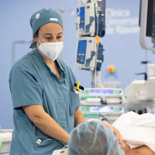
Our goal is to make your experience as comfortable and safe as possible. To achieve this, the entire interventional radiology team —including physicians, nurses, and technicians— will work in a coordinated manner, paying close attention to every detail and taking care of you at all times.
Sedation and Analgesia
To ensure your comfort and avoid discomfort, most procedures are performed under conscious sedation and local anesthesia at the puncture site. With this type of sedation, you remain relaxed but awake, allowing communication with the team at all times.
Throughout the procedure, your vital signs (pulse, blood pressure, oxygen saturation) will be continuously monitored to ensure maximum safety.
Average Duration
Although each case is different, the vast majority of interventional radiology procedures have an average duration ranging from 30 minutes to 2 hours. The medical team will always inform you of the estimated time for your specific procedure, adapted to your clinical situation.
Same-Day Discharge or Short Hospital Stay
One of the main advantages of these minimally invasive techniques is the rapid recovery they provide compared to open surgery.
In most cases, patients can be discharged the same day, after a short observation period in the recovery room to ensure everything is fine and to monitor the puncture site.
In more complex procedures, a short hospital stay may be necessary, usually around 24 hours, to provide closer monitoring before final discharge.

After the procedure
Care does not end when the procedure is finished. Follow-up is essential to ensure a complete recovery and to detect any issues in time.
Post-Discharge Care
When you return home, you will receive clear written instructions. In general, it is recommended to have relative rest during the first 24–48 hours, monitor the puncture site for possible bruising or bleeding, and maintain adequate hydration. It is essential to follow all instructions to prevent complications.
Warning Signs
Contact our team or go to the Emergency Department if you experience:
- Severe pain that does not improve with the prescribed pain medication.
- Significant bleeding or swelling at the puncture site.
- Fever or any other symptom that concerns you.
Team Follow-Up
Our commitment to you continues after discharge. We will schedule a follow-up, which may be by phone or in an in-person consultation, to assess your recovery, review the results of the procedure, and address any questions you may have.
For us, it is essential to ensure that the care you received has been optimal and has met your expectations.
Our team of professionals
TECHNOLOGY
Hybrid operating rooms
We have the best diagnostic imaging technology and state-of-the-art image-guided surgery, especially indicated for angiography and minimally invasive vascular interventions.
Ultrasound with image fusion
The ultrasound scanner with image fusion allows real-time combination of ultrasound images with other imaging modalities (CT, MRI, PET) facilitating biopsy and ablation procedures.


