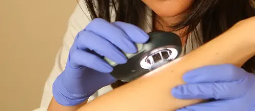
Superficial dermatophytic mycosis
"The treatments are very effective, but it is necessary to eliminate the circumstances that have caused it in order not to get it again".
DR. JAVIER ANTOÑANZAS
SPECIALIST. DERMATOLOGY DEPARTMENT

Mycoses are skin conditions resulting from parasitization by "fungi"; these are plants that do not carry out the phenomenon of photosynthesis.
They belong to the group of the most frequent diseases that affect man, and it can even be said that practically all men will suffer from them at some time in their lives.
There are three types of human mycosis: superficial, intermediate like candidiasis and deep. The usual ones in Spain are the superficial ones and the candidiasis.
The prognosis is good and they cure with treatment. It is important to remember that as long as the favorable circumstances are maintained, a new infection is likely.
It is also important to know that although they are not very contagious diseases they can be transmitted by direct contact with family members or indirectly with scales or hair or through combs, brushes, hats or towels.

What are the symptoms of mycosis?
The symptoms depend on the location, which in turn determines the classification of these mycoses.
- Tinea capitis.
- Tinea barbae.
- Tinea corporis.
- Tinea cruris.
- Tinea pedis.
- Tinea manum
- Onychomycosis or tinea ungium
The most common symptoms are:
- Flaky plaques on the hair.
- Bilateral plates of erythematous-brownish color with fine scales.
- Flaking in interdigital spaces.
Do you have any of these symptoms?
You may have a mycosis
What are the causes of mycosis?
They are caused by fungi belonging to the genera Epidermophyton, Microsporum and Trichophyton.
Who can suffer from mycosis?
Any person can sometimes have a dermatophytosis, which is facilitated by the existence of local predisposing factors such as moisture, occlusion and trauma. This explains why they are frequently located in the feet or in the groin region.
In addition, there are a series of general factors that also predispose to suffering from these mycoses, such as being subjected to general treatments with immunosuppressants or chemotherapy drugs, being diabetic, or suffering from ichthyosis or palm-plant keratoderma.
How are mycoses diagnosed?

The diagnosis of mycosis is usually made on a clinical basis.
Sometimes it is necessary to perform a direct examination of the skin scales or hairs to know if the causal agent is a dermatophyte or a yeast.
A culture of the lesions is necessary to determine the exact fungus responsible.
Depending on the clinical form and extent of the lesions, local or systemic treatment is indicated.
1. Tinea capitis. Affects scalp, eyebrows or eyelashes. There are two variants:
- Tinea capitis non-infammatory or dye tonsurantes. Preferably affects boys in second childhood causing real school epidemics. It can be manifested as one or few small scaly plaques where all the hair is cut a few millimeters. These plaques have a tendency to converge affecting large areas, or small plaques of alopecia may appear where it is possible to observe scales and healthy hair.
- Tinea capitis inflammatory (Celso's kerion): It is the most frequent in school children and preschoolers living in rural areas. It starts like the previous ones, but it gets indurated, it rises and it fills up with lesions of purulent content and flaky scabs that agglutinate the hair.
2. Tinea barbae. Preferably affects males in rural areas and is located in the area of the beard. It can be manifested as a reddish plate with small scales on its surface or as a red, edematous plate with pustules and covered by scabs.
3. Tinea corporis. It is located in the trunk, limbs and areas of the face without terminal hair. It can appear as circular or oval medallions with a scaly or vesicle edge and an erythematosquamous center or as a ring with a red edge and a cured center.
4. Tinea cruris. Also known as "marginal eczema of Hebra" it is located in English, perineum and perianal region being able to extend to internal proximal zone of thighs. Clinically it presents itself as bilateral plates of erythematous-brownish coloration with fine scales and a border of erythematous-vesiculous progression. It is important to remember that the infection is transmitted by towels, underwear and bedding. In men it can be associated with tinea pedis, since the fall of the fungus through the pants is quite frequent.
5. Tinea pedis. It is the most frequent ringworm since 15% of the people have suffered it or suffer it. It is known as "athlete's foot" and is located in interdigital spaces and soles of feet. Many times it is acquired by those who practice sports with bare feet or after playing go to showers for collective use. They are manifested as desquamation, maceration and cracking in interdigital spaces.
6. Tinea manum. It is located in folds, palm, back of hand being observed a descamative area in form of half moon.
7. Onychomycosis or Tinea ungium. The parasitation of the nail by "fungi" can be manifested as a thickening, detachment of the nail plate or as a change in color, acquiring a whitish hue.
How are mycoses treated?
Depending on the clinical form and extent of the lesions, local or systemic treatment is indicated.
Within the former, imidazole derivatives such as miconazole or ketoconazole are generally used. Terbinafine and amorolfine 5% are also effective.
Within the general treatments, griseofulvin, ketoconazole, itraconazole or terbinafine can be used.
In daily practice, these last two are frequently prescribed, with the exception of children who are prescribed griseofulvin.
The Department of Dermatology
of the Clínica Universidad de Navarra
The Department of Dermatology of the Clinica Universidad de Navarra has extensive experience in the diagnosis and treatment of dermatological diseases.
We have extensive experience in highly precise surgical treatments, such as Mohs surgery. This procedure requires highly specialized personnel.
We have the latest technology for the dermo-aesthetic treatment of skin lesions, with the aim of achieving the best results for our patients.
Diseases we treat

Why at the Clinica?
- Experts in Mohs Surgery for the treatment of skin cancer.
- We have the best technology for dermo-aesthetic treatments.
- Safety and quality assurance of the best private hospital in Spain.


