Cardiac Imaging
Biomedical Imaging Laboratory
Myocardial perfusion
Myocardial Blood Flow (MBF) is an important index of ischemic heart disease. First pass perfusion MRI permits rapid measurement of MBF, but requires the use of a gadolinium-based contrast agent. Despite the fact that this is a commonly used technique, the need of injecting Gadolinium prevents its use in patients with renal impairment and limits the repeatability of the technique.
Within this context, the goal is to develop a new imaging technique to quantify the delivery of blood to the myocardial tissue (perfusion) and to translate this technique from healthy to patient-oriented studies.

Do you need us to help you?
Contact us and request more information.
Myocardial arterial spin labeling (ASL)
Arterial spin labeling (ASL) offers a noninvasive and repeatable alternative for non-contrast perfusion measurements.
It does not require injection of a contrast agent due to the fact that it uses blood as an endogenous tracer. In order to label blood entering the coronary arteries of the heart, FAIR labeling is used.
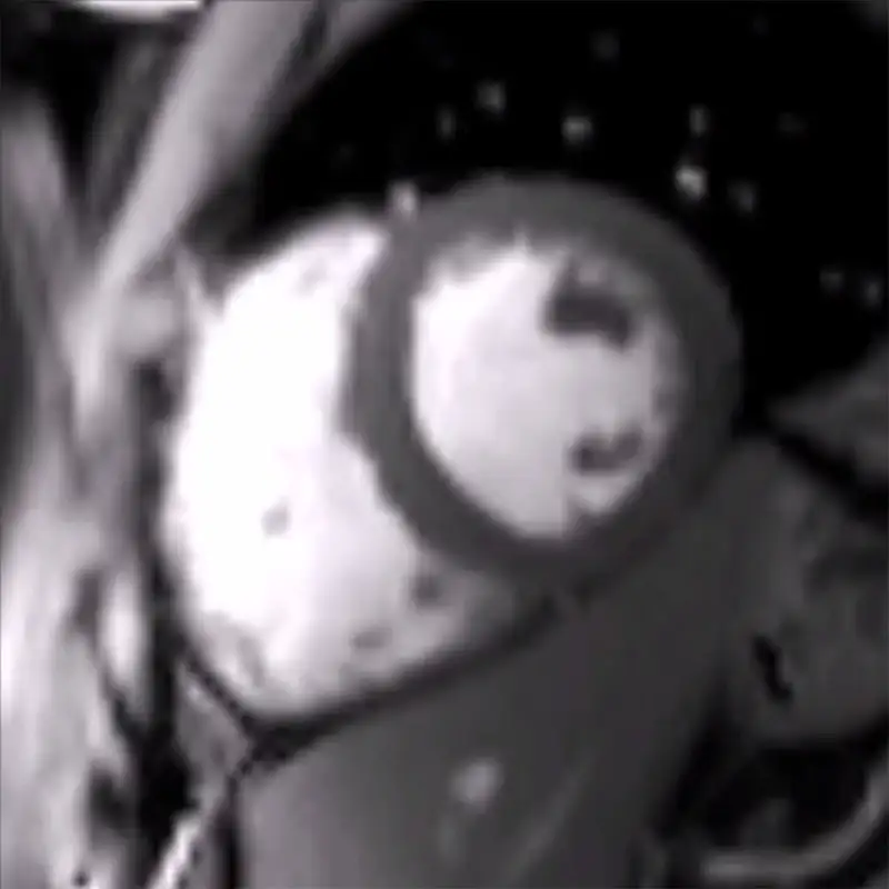
Here, global and selective magnetic inversions are alternated, followed by a delay period (of approximately 1 second) to allow labeled blood flow into the myocardial tissue before the MRI image is acquired. The global inversion image (label) inverts both static tissue and blood. The selective inversion image (control) only inverts the static tissue.
Subtraction of both images gives information of the delivery of blood to the myocardial tissue.
Figure 1. On the left, global inversion (label). On the right, selective inversion (control).
To increase SNR, several images need to be acquired during a cardiac study. This can be performed during free breathing. Subsequently, images are coregistered to minimize the effects of respiratory motion.
The following videos show the original acquired images and the subsequently registered images. Registration has been performed using a non-rigid transformation, which allows a wide variety of deformations.

After image acquisition, pairwise subtraction of label of control images is performed to obtain perfusion-weighted images. These images are then averaged. Finally, a general kinetic model equation is used to quantify myocardial blood flow.
The following image shows the MBF map of a healthy volunteer at rest. The colors represent quantitative units in milliliters of blood per gram of tissue per minute (ml/g/min).

Figure 2. On the left, anatomic reference image. On the right, quantitative MBF map obtained with ASL sequence in ml/g/min at rest.
In order to simulate a similar perfusion increase to that obtained during pharmacological stress conditions, a passive leg elevation task was performed with healthy volunteers. A wooden box with 50º elevation was used and the task started 5 minutes before scanning.

Figure 3. Wooden box with 50º elevation used to simulate stress conditions and increase myocardial perfusion in a healthy volunteer.
The following figure shows the MBF maps of a representative healthy volunteer obtained at rest and during mild stress conditions with passive leg raising.

Figure 4. On the left, myocardial blood flow map obtained at rest. On the right, myocardial blood flow map obtained during mild stress with passive leg raising in a healthy volunteer.
Our team of professionals


